Home » Without Label » Bones In Leg Diagram - Human Leg Bones PNG Images & PSDs for Download ... - The hip itself is a ball and socket joint, much like the shoulder.the structures necessary to create this joint are the socket, the joint capsule, muscle, ligaments, and the neck.
Bones In Leg Diagram - Human Leg Bones PNG Images & PSDs for Download ... - The hip itself is a ball and socket joint, much like the shoulder.the structures necessary to create this joint are the socket, the joint capsule, muscle, ligaments, and the neck.
Bones In Leg Diagram - Human Leg Bones PNG Images & PSDs for Download ... - The hip itself is a ball and socket joint, much like the shoulder.the structures necessary to create this joint are the socket, the joint capsule, muscle, ligaments, and the neck.. The lower leg extends from the knee to the ankle. (note, the radius and ulna bones also have this membrane.) this membrane keeps the tibia and fibula together and provides strength and stability for them. In the leg muscles diagram above, there are many muscles that make up your legs and support it to move. Also called the shin bone, the tibia is the. The stifle joint connects the femur, which is the dog thigh bone, to the tibia and fibula, the lower leg bones, and the patella,the canine equivalent to the knee cap.
Like the upper limb, the lower limb is divided into three regions. Femur, patella, tibia, fibula—these words may sound super scientific, but they're just the names of the bones in our legs! Tibia and fibula the tibia and fibula are two long bones that run parallel to each other, forming the scaffold of the leg and providing attachment points for many muscles. The tibia and fibula form the ankle joint with the talus,. The lower leg extends from the knee to the ankle.
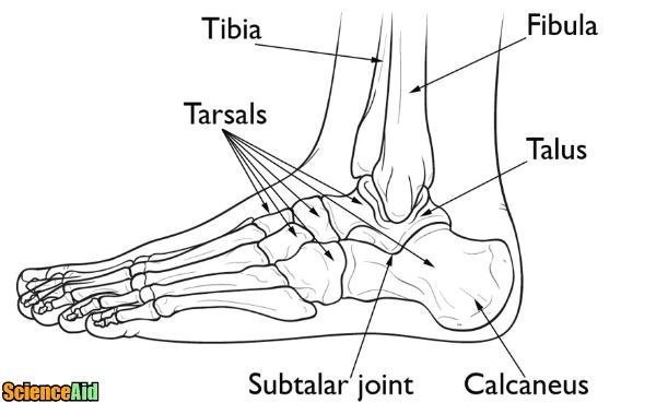
Bones of the Human Leg and Foot - ScienceAid from scienceaid.net In the leg muscles diagram above, there are many muscles that make up your legs and support it to move. It also separates muscles on the anterior and posterior parts of the leg. These muscles work together to produce movements such as standing, walking, running, and jumping. Degenerative disease, similar to arthritis. These are the femur, patella, tibia, fibula, tarsal bones, metatarsal bones, and phalanges (see. Bone diagram forehead (frontal bone) nose bones (nasals) cheek bone (zygoma) upper jaw (maxilla) lower jaw (mandible) breast bone (sternum) upper arm bone (humerus) lower arm bone (ulna) thigh bone (femur) collar bone (clavicle) toe bones (phalanges) ankle bones (tarsals) kneecap (patella) shin bone Numbered one through five the bone that sits behind the big toe is no. The tibia, commonly known as the 'shin bone', is the largest and most medial of the two.you can palpate its anterior border when you run your finger down the anterior aspect of your leg.
This is the first joint in the leg.
The tibia, commonly known as the 'shin bone', is the largest and most medial of the two.you can palpate its anterior border when you run your finger down the anterior aspect of your leg. Numerous bone is the long bone of the upper arm which goes all the way to the elbow. Ulna and the radius are two bones that sit next to each other. They are numbered from one to five, starting from the medial (inner) side of the foot. A muscle strain is an injury to a muscle or a tendon — the fibrous tissue that connects muscles to bones. Its lower end helps create the knee joint. These are the femur, patella, tibia, fibula, tarsal bones, metatarsal bones, and phalanges (see. The lower leg lies between the knee and the ankle. It is made of the ulna and the radius. It is likely that abnormal biomechanical stresses are the basis for the disease. The lower leg is comprised of two bones the tibia and the smaller fibula. Related posts of diagram of leg bones inside of arm muscle and bone. At the same time, the bones and joints of the leg and foot must be strong enough to support the body's weight while remaining.
The technical term for a dog knee is the stifle joint. This diagram depicts diagram leg bones anatomy. It also separates muscles on the anterior and posterior parts of the leg. Inside of arm muscle and bone 12 photos of the inside of arm muscle and bone , bone Tibia and fibula the tibia and fibula are two long bones that run parallel to each other, forming the scaffold of the leg and providing attachment points for many muscles.
Leg Bones - Medical Art Library from www.medicalartlibrary.com There are three bones in the knee namely the femur which is the thigh bone, tibia which is the shin bone and patella which is the knee cap. Leg bone anatomy diagram diagram of human leg human anatomy human leg bones anatomy stock photo download image now anatomy of the knee central coast orthopedic medical group This is the first joint in the leg. Like the upper limb, the lower limb is divided into three regions. A muscle strain is an injury to a muscle or a tendon — the fibrous tissue that connects muscles to bones. Dog leg anatomy is complex, especially dog knees, which are found on the hind legs. The lower leg is comprised of two bones the tibia and the smaller fibula. Jul 16, 2019 · the bones of the leg and foot form part of the appendicular skeleton that supports the many muscles of the lower limbs.
The given diagram of the knee joint can help you to understand its various parts and the description given below will give you an insight of the functioning of the knee.
The thigh and leg bones articulate at the knee joint that is protected and enhanced by the patella bone that supports the quadriceps tendon. Related posts of diagram of leg bones inside of arm muscle and bone. Numbered one through five the bone that sits behind the big toe is no. The fibula is mainly a muscle attachment point and is used to help maintain balance. Tibia and fibula the tibia and fibula are two long bones that run parallel to each other, forming the scaffold of the leg and providing attachment points for many muscles. Continue scrolling to read more below. The lower leg extends from the knee to the ankle. Ulna and the radius are two bones that sit next to each other. In the leg muscles diagram above, there are many muscles that make up your legs and support it to move. Now let's look at the tibia bone, which is the larger of the two leg bones, located medially. The femur, or thighbone, is the longest and largest bone in the human body. Also called the shin bone, the tibia is the. It is likely that abnormal biomechanical stresses are the basis for the disease.
The bones of the hip include the femur, the ilium, the ischium, and the pubis. Now let's look at the tibia bone, which is the larger of the two leg bones, located medially. In the leg muscles diagram above, there are many muscles that make up your legs and support it to move. The thigh bone, or femur, is the large upper leg bone that connects the lower leg bones (knee joint) to the pelvic bone (hip joint). Now i will provide you the few information on the other bones of dog leg anatomy with their unique features.
Infographic Diagram Of Human Femur Bone Or Leg Bone ... from media.istockphoto.com The femur, or thighbone, is the longest and largest bone in the human body. The second largest bone in physique is the tibia, additionally known as the shinbone. The elbow is located below the chest at the back of the foreleg. The pubis, ischium, and ilium together constitute the pelvis while the thigh bone is the femur. At the same time, the bones and joints of the leg and foot must be strong enough to support the body's weight while remaining. The tibia, commonly known as the 'shin bone', is the largest and most medial of the two.you can palpate its anterior border when you run your finger down the anterior aspect of your leg. Now i will provide you the few information on the other bones of dog leg anatomy with their unique features. The thigh and leg bones articulate at the knee joint that is protected and enhanced by the patella bone that supports the quadriceps tendon.
Another bone that is part of the lower leg and the knee joint is called the fibula.this is a bone located on the lateral, or outer part, of the lower leg and is more commonly known as the calf bone.
Muscle spasms, cramps and injuries can all cause muscle pain. Performance horses tend to suffer from this degenerative disease. Schema de legs bones diagram diagram showing bones inside human leg ready to jump stock file skeleton of a cat diagram ver 2 svg disposition of rotator cuff muscles diagram. Bone diagram forehead (frontal bone) nose bones (nasals) cheek bone (zygoma) upper jaw (maxilla) lower jaw (mandible) breast bone (sternum) upper arm bone (humerus) lower arm bone (ulna) thigh bone (femur) collar bone (clavicle) toe bones (phalanges) ankle bones (tarsals) kneecap (patella) shin bone The tibia and fibula are the bones of the lower leg. The front leg of a dog consists of the clavicle, scapula (arm), radius and ulna (forearm), carpals, metacarpals, and phalanges (forepaw). This diagram depicts diagram leg bones anatomy. These are the femur, patella, tibia, fibula, tarsal bones, metatarsal bones, and phalanges (see. The tibia, commonly known as the 'shin bone', is the largest and most medial of the two.you can palpate its anterior border when you run your finger down the anterior aspect of your leg. Inflammation of navicular bone and/or bursa. The bones of your leg have roughened patches on their surfaces where muscles are attached. Related posts of diagram of leg bones inside of arm muscle and bone. Bone fracture human leg anatomy and skeleton stock vector leg bones diagram diagram schematic ideas diagram showing bones inside human leg ready to jump stock explore more like human leg bones diagram.